
Diagnostic imaging, often shortened in medical contexts to “diag image,” is a revolutionary field of medicine that allows healthcare professionals to view the internal structures of the human body non-invasively. It serves as a critical decision-making tool, moving beyond educated guesses to provide visual evidence of disease, injury, or abnormality. The core principle of diagnostic imaging is to capture detailed pictures of our internal anatomy and physiological processes, providing a window into the body’s most hidden secrets. This article explores the various modalities of diagnostic imaging, their applications, and their indispensable role in modern healthcare.
The Pillars of Modern Medicine: Key Imaging Modalities
Diagnostic imaging is not a single technology but a suite of complementary techniques, each with unique strengths. The most common modalities include:
- X-Ray (Radiography): The oldest and most widely used form of medical imaging. X-rays work by passing a small amount of ionizing radiation through the body. Dense structures like bones absorb the radiation and appear white on the resulting image, while softer tissues appear in shades of gray. X-rays are typically the first-line investigation for fractures, dental issues, and chest infections like pneumonia.
- Computed Tomography (CT or CAT Scan): A CT scan combines a series of X-ray images taken from different angles around the body. A computer then processes these images to create cross-sectional, detailed slices of bones, blood vessels, and soft tissues. CT scans provide far more detail than a standard X-ray and are invaluable for diagnosing cancers, locating tumors, identifying internal bleeding, and examining complex fractures.
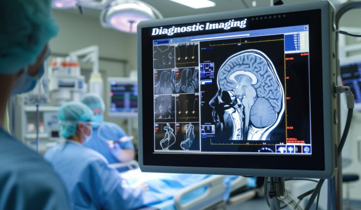
- Magnetic Resonance Imaging (MRI): Instead of radiation, an MRI uses powerful magnets and radio waves to generate highly detailed images of organs and soft tissues. It is exceptionally effective for visualizing the brain, spinal cord, nerves, muscles, ligaments, and tendons. MRI is the preferred modality for diagnosing neurological conditions (like multiple sclerosis), joint injuries, and many musculoskeletal problems due to its superior contrast resolution for soft tissues.
- Ultrasound (Sonography): This modality uses high-frequency sound waves to produce real-time images, known as sonograms, of the inside of the body. Because it does not use ionizing radiation, it is the go-to technique for monitoring fetal development during pregnancy. It is also commonly used to examine abdominal organs, the heart (echocardiogram), breasts, and blood flow through vessels (Doppler ultrasound).
- Nuclear Medicine (PET & SPECT Scans): These advanced techniques involve administering a small amount of radioactive tracer into the patient’s body. This tracer builds up in particular tissues or organs. Special cameras then detect the radioactivity and create images that show both structure and, uniquely, function. Positron Emission Tomography (PET) scans, for instance, are crucial in oncology for detecting cancer metastasis and measuring cellular metabolic activity.
The Integral Role of the Medical Imaging Workstation
The acquisition of the image is only the first step. The captured data is sent to a dedicated medical imaging workstation. These are high-performance computers equipped with specialized software and high-resolution displays calibrated for medical use. Radiologists—physicians trained to interpret these images—use these workstations to:
- Manipulate Images: They can adjust contrast, zoom in on areas of interest, and take precise measurements.
- Create 3D Reconstructions: For CT and MRI scans, powerful software can compile the 2D slices into intricate 3D models, providing a comprehensive view of complex structures like the vascular system or a tumor’s relationship to surrounding organs.
- Compare Studies: Radiologists can compare new images with a patient’s prior scans to track the progression of a disease or the effectiveness of a treatment over time.
- Generate Reports: Their detailed interpretations are compiled into formal reports that are sent to the referring physician, who uses this information to make a final diagnosis and determine the best course of treatment.
The Impact on Diagnostic Accuracy and Patient Outcomes
The value of diagnostic imaging is profound. By providing a clear internal view, it:
- Enables Early and Accurate Diagnosis: Many serious conditions, including cancers, strokes, and heart disease, have vastly better outcomes when detected early. Imaging allows for identification long before physical symptoms may become severe.
- Guides Treatment: Imaging is essential for planning surgeries, biopsies, and radiation therapy. It acts as a map for surgeons, allowing for precision and minimizing damage to healthy tissues.
- Reduces the Need for Exploratory Surgery: Before these technologies were available, surgeons often had to operate simply to diagnose a problem. Now, non-invasive imaging can provide a definitive answer, saving patients from unnecessary procedures and risks.
- Monitors Treatment Efficacy: Repeat scans during a treatment regimen (e.g., chemotherapy) show clinicians whether a tumor is shrinking, allowing them to adjust therapies that aren’t working.
While studies indicate that diagnostic errors, including those related to imaging interpretation, still occur, advancements in technology are directly aimed at reducing them. Artificial Intelligence (AI) is now being integrated into imaging workstations to act as a “second set of eyes,” using algorithms to flag potential abnormalities a human might miss, further enhancing diagnostic precision.
Is Imaging Always Necessary?
A common patient concern is exposure to radiation from certain scans like X-rays and CTs. It is important to understand that physicians follow the principle of ALARA (As Low As Reasonably Achievable). They will only order an imaging test when the clinical benefits significantly outweigh the minimal risks. Your doctor will consider your specific symptoms, medical history, and other factors to determine if an imaging study is the most appropriate next step. It is a powerful tool, but one used judiciously to guide effective treatment and ultimately, restore health.
Frequently Asked Questions (FAQs)
Q1: What is the difference between a CT scan and an MRI?
A: The key difference is the technology used. A CT scan uses X-rays and is best for viewing bony structures, bleeding, and trauma. An MRI uses magnets and radio waves and is superior for imaging soft tissues like the brain, muscles, and ligaments. The choice depends on what part of the body needs to be examined.
Q2: Are medical imaging procedures painful?
A: Most diagnostic imaging procedures are painless. The discomfort, if any, usually comes from having to remain still for an extended period on a hard table. Some procedures, like certain MRI scans, may involve an injection of a contrast agent, which can feel like a brief pinprick.
Q3: How should I prepare for an imaging appointment?
A: Preparation varies greatly by the type of scan. You may be asked to fast (not eat or drink) for several hours before an abdominal ultrasound or CT scan. For an MRI, you will be screened for any metal implants in your body. Always follow the specific instructions provided by your healthcare provider and the imaging center.
Q4: Who interprets my imaging results?
A: A specialized doctor called a radiologist interprets your scans. They are experts in analyzing medical images. They then send a detailed report to the doctor who ordered the test (e.g., your primary care physician or surgeon), who will discuss the results and their meaning with you.
Q5: Is the radiation from X-rays and CT scans dangerous?
A: The radiation doses from diagnostic imaging are very low and are considered safe for adults. The medical benefit of getting an accurate diagnosis almost always far outweighs the minimal risk from radiation exposure. Doctors are careful to avoid unnecessary imaging, especially in pregnant women and children.